Foot Muscles Mri : МРТ стопы | vsemrt.ru : Flexion of 4 lesser toes at metatarsophalangeal, proximal & distal interphalangeal joints inversion of foot plantar flexion of ankle.
byAdmin•
0
Foot Muscles Mri : МРТ стопы | vsemrt.ru : Flexion of 4 lesser toes at metatarsophalangeal, proximal & distal interphalangeal joints inversion of foot plantar flexion of ankle.. Routine ankle magnetic resonance imaging (mri) tests involve taking images of the foot and ankle in the axial, coronal, and sagittal planes the imaging process allows the magnetic field to find changes in the organ and tissue structures, identifying any sprains, ruptures, dislocations, or synovial disorders. This is the first of two parts on the intrinsic muscles of the foot. This is a 30 year old with swelling on the lateral aspect of foot with evidence of soft tissue lesion in relation to the lateral aspect of the talus which appears isointense to the muscles on t1 and t2 weighted images & appears elongated extending from the anterosuperior calcaneum to the base of. Indications for foot mri scan. Bone contusions, osteonecrosis, marrow oedema syndromes, and stress > fractures) > synovial based disorders ( e.g.
It begins with short tendon bundles on the medial surface of the calcaneus calcaneus, fleshy bundles on the lower retentive flexor. Human anatomy for muscle, reproductive, and skeleton. This means that the little toe can only be extended by the extensor digitorum longus muscle only. The muscles acting on the foot can be divided into two distinct groups; The muscles of the dorsum of the foot are a group of two muscles, which together represent the dorsal foot musculature.
Foot radiological anatomy. shorouk zaki from image.slidesharecdn.com Routine ankle magnetic resonance imaging (mri) tests involve taking images of the foot and ankle in the axial, coronal, and sagittal planes the imaging process allows the magnetic field to find changes in the organ and tissue structures, identifying any sprains, ruptures, dislocations, or synovial disorders. Mri with hardware in foot? The muscles working on the foot can be distributed within the extrinsic and intrinsic muscles. Bone contusions, osteonecrosis, marrow oedema syndromes, and stress > fractures) > synovial based disorders ( e.g. For instance, i am having an mri of my foot next week, and have to remove all jewellry. Perform routine foot plus coronal fmpspgr fat saturated pre and post gad images and axial post gad fmpspgr fat saturated images. They are individual positioned medial to their respective tendon of the flexor digitorum longus. Mr data were then acquired.
► shoulder ► elbow ► wrist ► finger ► thumb.
Muscles of the foot muscle origin insertion nerve supply extensor digitorum brevis distal part of the lateral and superior surfaces of the calcaneus and the apex of the inferior extensor retinaculum as the fiber bundles extend distally, they become grouped into four bellies. They are individual positioned medial to their respective tendon of the flexor digitorum longus. Muscles of the ankle and foot. Perform routine foot plus coronal fmpspgr fat saturated pre and post gad images and axial post gad fmpspgr fat saturated images. Involved early gray = muscle: As a result, during walking the body's center of gravity normally fluctuates only 5cm in both vertical and lateral directions. Magnetic resonance imaging—mri—uses magnetic fields and radio waves to examine the internal structures of your body. Synovitis, tenosynovitis, bursitis, and ganglion cysts) > congenital and developmental conditions ( eg.dysplasia. This is a 30 year old with swelling on the lateral aspect of foot with evidence of soft tissue lesion in relation to the lateral aspect of the talus which appears isointense to the muscles on t1 and t2 weighted images & appears elongated extending from the anterosuperior calcaneum to the base of. The purpose of this study was to investigate the relationship of muscle mri findings and gait disturbance in myotonic dystrophy type 1 (dm1) patients. A magnetic resonance imaging (mri) was performed on a normal subject; Bone contusions, osteonecrosis, marrow oedema syndromes, and stress > fractures) > synovial based disorders ( e.g. Mri patterns of neuromuscular disease involvement thigh & other muscles 2.
A magnetic resonance imaging (mri) was performed on a normal subject; The muscles acting on the foot can be divided into two distinct groups; Indications for foot mri scan. An overview of the intrinsic muscles of the foot including their origin, insertion, blood supply, innervation, function and clinical relevance. Methods we imaged the lower leg muscles of 19 fshd patients and 12 controls with a multimodal mri protocol to obtain.
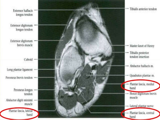
Muscles of the Foot - Dorsal - Plantar - TeachMeAnatomy from teachmeanatomy.info The abductor digiti minimi muscle is on the lateral side of the foot and contributes to the large lateral plantar eminence on the sole. This article reviews the use of magnetic resonance imaging (mri) in the evaluation of the foot, including a discussion of bone and cartilage abnormalities in an article published in the august 2006 issue of this journal, the authors reviewed magnetic resonance imaging (mri) of the ankle. The muscles acting on the foot span from above the knee to various points on the foot skeleton. Synovitis, tenosynovitis, bursitis, and ganglion cysts) > congenital and developmental conditions ( eg.dysplasia. Flexion of 4 lesser toes at metatarsophalangeal, proximal & distal interphalangeal joints inversion of foot plantar flexion of ankle. Top suggestions for foot muscle anatomy mri. Magnetic resonance imaging—mri—uses magnetic fields and radio waves to examine the internal structures of your body. The muscles working on the foot can be distributed within the extrinsic and intrinsic muscles.
Muscles of the ankle and foot.
These muscles lengthen eccentrically during the stance phase of running before shortening at the propulsion phase. Muscle strength) for the foot dorsal and plantar flexors 23. Human anatomy for muscle, reproductive, and skeleton. Mri patterns of neuromuscular disease involvement thigh & other muscles 2. The muscle that removes the big toe (m.abductor hallucis) lies superficially along the medial edge of the foot. This is a 30 year old with swelling on the lateral aspect of foot with evidence of soft tissue lesion in relation to the lateral aspect of the talus which appears isointense to the muscles on t1 and t2 weighted images & appears elongated extending from the anterosuperior calcaneum to the base of. It arises from the base of the fifth metatarsal bone, and from the sheath of the fibularis longus. Perform routine foot plus coronal fmpspgr fat saturated pre and post gad images and axial post gad fmpspgr fat saturated images. Related posts of foot muscle anatomy mri. The flexor digiti minimi brevis (flexor brevis minimi digiti, flexor digiti quinti brevis) lies under the metatarsal bone on the little toe, and resembles one of the interossei. The instructions also say no hair spray/mousse/gel etc. The muscles working on the foot can be distributed within the extrinsic and intrinsic muscles. It begins with short tendon bundles on the medial surface of the calcaneus calcaneus, fleshy bundles on the lower retentive flexor.
The purpose of this study was to investigate the relationship of muscle mri findings and gait disturbance in myotonic dystrophy type 1 (dm1) patients. Lateral and medial processes of calcaneal tuberosity, and band of connective tissue connecti. There can't be any metal in the room, not just where you have the mri. Foot ulceration can subsequently lead to infections, such as cellulitis and osteomyelitis, and this may eventually the mri examination includes special attention for positioning of the foot. Involved early gray = muscle:
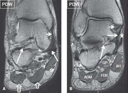
Plantar Foot Muscles Mri / Central plantar muscles of the ... from musculoskeletalkey.com Routine ankle magnetic resonance imaging (mri) tests involve taking images of the foot and ankle in the axial, coronal, and sagittal planes the imaging process allows the magnetic field to find changes in the organ and tissue structures, identifying any sprains, ruptures, dislocations, or synovial disorders. The muscles of the dorsum of the foot are a group of two muscles, which together represent the dorsal foot musculature. Interestingly the dorsal foot muscles generally have no insertion at the little toe. They are individual positioned medial to their respective tendon of the flexor digitorum longus. This is the first of two parts on the intrinsic muscles of the foot. Evaluated the energy reserves in foot muscles using mri measurements of phosphorus metabolites. There are 10 intrinsic muscles located in the sole of the foot. An overview of the intrinsic muscles of the foot including their origin, insertion, blood supply, innervation, function and clinical relevance.
Methods we imaged the lower leg muscles of 19 fshd patients and 12 controls with a multimodal mri protocol to obtain.
It must be placed in the center of the magnet, to obtain homogeneous fat. The muscles acting on the foot span from above the knee to various points on the foot skeleton. Human anatomy for muscle, reproductive, and skeleton. Indications for foot mri scan. Evaluated the energy reserves in foot muscles using mri measurements of phosphorus metabolites. Mr data were then acquired. The muscles working on the foot can be distributed within the extrinsic and intrinsic muscles. This means that the little toe can only be extended by the extensor digitorum longus muscle only. It begins with short tendon bundles on the medial surface of the calcaneus calcaneus, fleshy bundles on the lower retentive flexor. They act collectively to stabilise the arches of the foot, and individually to control movement of the digits. This is the first of two parts on the intrinsic muscles of the foot. There are 10 intrinsic muscles located in the sole of the foot. The muscle that removes the big toe (m.abductor hallucis) lies superficially along the medial edge of the foot.